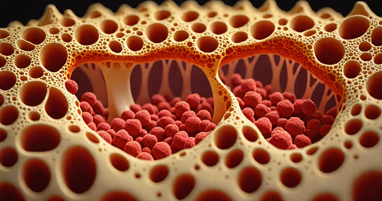
Bone marrow formation is the biological process by which hematopoietic stem cells in the bone marrow differentiate into all types of blood cells-red blood cells, white blood cells, and platelets-through a tightly regulated sequence of cellular divisions and specialization. This continuous production sustains oxygen transport, immune defense, and clotting ability throughout life.
The process begins in the fetal stage, primarily in the liver and spleen, then shifts to the bone marrow by the third trimester. After birth, the bone marrow becomes the central site for blood cell generation. By age 20, nearly all hematopoiesis occurs in the marrow cavities of long bones (like the femur and humerus), flat bones (such as the pelvis and sternum), and vertebrae.
Each day, the human body produces approximately 200 billion red blood cells, 100 billion platelets, and tens of billions of white blood cells-all originating from a single multipotent hematopoietic stem cell (HSC). These HSCs reside in specialized microenvironments called niches within the marrow stroma, where signals from surrounding cells regulate their quiescence, activation, and lineage commitment.
Bone marrow formation follows a hierarchical model. Hematopoietic stem cells give rise to multipotent progenitors, which then commit to specific lineages-myeloid or lymphoid. Myeloid lineage leads to granulocytes, monocytes, erythrocytes, and megakaryocytes. Lymphoid lineage produces B cells, T cells, and natural killer cells.
Cell differentiation is guided by cytokines and growth factors. For example, erythropoietin (EPO) stimulates red blood cell production in response to low oxygen levels. Granulocyte colony-stimulating factor (G-CSF) boosts neutrophil counts during infection. These signals act on precursor cells in the marrow, triggering proliferation and maturation over several days.
A key feature is the balance between self-renewal and differentiation. A stem cell must divide to maintain its pool while also producing daughter cells that mature into functional blood cells. Disruption of this balance leads to conditions such as leukemia (excessive immature cells) or aplastic anemia (failure to produce enough cells).
Bone marrow contains two primary types of tissue: red marrow and yellow marrow. Red marrow is metabolically active and responsible for blood cell formation. Yellow marrow consists mainly of fat cells and is inactive in hematopoiesis. In adults, red marrow occupies the central cavities of the pelvis, sternum, skull, ribs, and vertebrae.
| Type | Primary Function | Location in Adult Body | Cell Yield |
|---|---|---|---|
| Red marrow | Active hematopoiesis | Pelvis, sternum, vertebrae, proximal femur and humerus | Produces all blood cell types |
| Yellow marrow | Fat storage, limited hematopoiesis | Distal long bones (femur, tibia) | Minimal blood cell output |
When blood loss or infection occurs, the body can reactivate dormant yellow marrow sites to resume blood cell production-a phenomenon known as extramedullary hematopoiesis. This rarely happens in healthy adults but may occur in severe anemia or chronic disease.
An often overlooked fact: bone marrow formation is not uniform across the skeleton. The pelvis generates more blood cells per gram of marrow than the femur. This regional variation reflects differences in vascular density, oxygen tension, and niche signaling molecules.
Disorders of bone marrow formation underlie many serious diseases. Aplastic anemia results from damage to HSCs, leading to pancytopenia-low counts of all blood cell types. Leukemias arise from mutations in early progenitor cells, causing uncontrolled proliferation of abnormal white blood cells. Multiple myeloma involves malignant plasma cells that crowd out normal marrow elements.
Diagnosis relies on bone marrow aspiration and biopsy. A sample is drawn from the posterior iliac crest using a needle. Pathologists examine the cellular composition, assess cellularity, and detect abnormal morphology. Normal marrow has 30-70% cellularity, with balanced proportions of erythroid, myeloid, and megakaryocytic precursors.
I once observed a 42-year-old patient with fatigue and petechiae. His CBC showed severe thrombocytopenia and normocytic anemia. A bone marrow biopsy revealed hypocellularity and absence of megakaryocytes-diagnostic of idiopathic aplastic anemia. Treatment involved immunosuppressive therapy and stem cell transplantation. The patient's marrow recovery took six months, highlighting how slow and delicate the restoration of bone marrow formation can be.
Bone marrow formation is frequently confused with blood cell aging or destruction. The spleen and liver clear old red blood cells, but they do not produce new ones. Bone marrow is the sole site of ongoing hematopoiesis in adults.
Another misconception is that bone marrow forms only red blood cells. In reality, it produces all major blood cell types. Platelets derive from megakaryocytes, which fragment into thousands of small particles. Neutrophils and lymphocytes originate from myeloid and lymphoid progenitors, respectively.
Some assume that bone marrow is static. It is not. Marrow activity increases during stress-such as hemorrhage, infection, or chemotherapy-by releasing stored progenitors and expanding niche capacity. This adaptability allows rapid recovery after injury.
Bone marrow formation depends on a supportive stromal environment. Mesenchymal stem cells secrete factors like SCF (stem cell factor) and CXCL12, which anchor HSCs and promote survival. Disruption of these signals impairs hematopoiesis. For example, high-dose radiation damages stromal cells, leading to prolonged cytopenias.
Understanding how bone marrow is produced involves tracing the developmental pathway from embryonic mesoderm to adult hematopoietic tissue. The process begins around week 6 of gestation, with the emergence of hemangioblasts in the yolk sac. By week 8, definitive hematopoiesis starts in the aorta-gonad-mesonephros (AGM) region. The fetal liver takes over until the bone marrow becomes dominant.
Bone marrow formation is not a single event-it is a lifelong, changing process essential to human health. Its failure disrupts oxygen delivery, immunity, and hemostasis. Its resilience enables recovery from illness and injury. This complex system, operating beneath the surface of the skeleton, maintains the body's most fundamental circulatory functions.