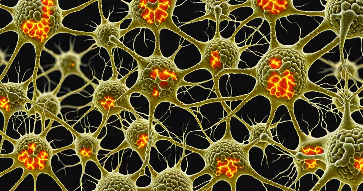
Neurons in the brain are specialized nerve cells that process and transmit information via electrical and chemical signals, forming the biological basis of thought, emotion, movement, and perception. They communicate through synapses, creating vast interconnected networks essential for all brain functions.
Each neuron consists of a cell body (soma), dendrites that receive signals, and an axon that sends signals to other neurons or muscles. When stimulated, a neuron fires an electrical impulse called an action potential, which travels down the axon and triggers neurotransmitter release at the synapse. This process enables communication across billions of connections.
The human brain contains approximately 86 billion neurons, organized into distinct regions that control specific functions. For example, motor neurons in the cerebral cortex initiate voluntary movements, while sensory neurons in the occipital lobe process visual input. These cells do not operate in isolation-they form changing circuits that adapt based on experience, learning, and injury.
Neurons function through electrochemical signaling. A resting neuron maintains a negative internal charge (about -70 millivolts) due to ion distribution across its membrane. When a stimulus reaches the dendrites, it alters the membrane potential. If the threshold (around -55 mV) is crossed, voltage-gated sodium channels open, triggering an action potential that propagates along the axon.
At the axon terminal, the electrical signal causes vesicles filled with neurotransmitters-such as dopamine, serotonin, or glutamate-to fuse with the membrane and release their contents into the synaptic cleft. These chemicals bind to receptors on the receiving neuron's dendrites, either exciting or inhibiting its activity. This process occurs in milliseconds and underlies everything from reflexes to complex decision-making.
A single neuron can connect to thousands of others. The hippocampus, involved in memory formation, contains neurons that fire in synchronized bursts during learning. Research using functional MRI shows that during a memory task, specific neuronal ensembles activate in sequence, reinforcing pathways associated with recall.
Neurons vary by shape, size, and function. The three main categories are sensory neurons, motor neurons, and interneurons. Sensory neurons carry signals from the body to the central nervous system, such as pain or touch sensations. Motor neurons send commands from the brain to muscles, enabling movement. Interneurons, which make up about 99% of neurons in the brain, connect other neurons within the CNS and are critical for integrating information.
| Type | Function | Location | Example |
|---|---|---|---|
| Sensory | Transmit external stimuli | Spinal cord, peripheral nerves | Touch receptors in fingertips |
| Motor | Control muscle contraction | Cerebral cortex, spinal cord | Neurons activating leg muscles during walking |
| Interneuron | Process and integrate signals | Brain gray matter | Hippocampal neurons encoding episodic memories |
Neurons also differ in morphology. Pyramidal neurons in the cerebral cortex have a triangular-shaped soma and long axons that project to distant areas. Purkinje cells in the cerebellum have elaborate dendritic trees shaped like fans, allowing them to receive input from tens of thousands of other neurons. These structural differences support specialized roles in information processing.
Neurons in the brain enable cognitive functions such as attention, problem-solving, emotional regulation, and language. Damage to specific populations leads to measurable deficits. For instance, degeneration of dopaminergic neurons in the substantia nigra causes Parkinson's disease, characterized by tremors and slowed movement. Loss of neurons in the hippocampus correlates with memory loss in Alzheimer's disease.
Neuroplasticity-the brain's ability to reorganize itself-relies on neurons adapting their connections. After a stroke, surviving neurons can take over lost functions by strengthening existing synapses or forming new ones. This adaptation occurs through mechanisms like long-term potentiation (LTP), where repeated stimulation increases synaptic strength. Studies show that LTP can last for weeks in animal models, supporting lasting changes in neural circuits.
One non-obvious fact: neurons in the adult human brain do not divide. Unlike skin or liver cells, they don't regenerate through mitosis. Instead, new neurons arise from neural stem cells in limited regions-primarily the subventricular zone and the dentate gyrus of the hippocampus. This process, called neurogenesis, was confirmed in humans using carbon-14 dating of neuron DNA, which showed that some hippocampal neurons are less than 10 years old. This challenges the outdated idea that the adult brain cannot grow new cells.
Many assume that "thinking" happens in a single brain region. In reality, thoughts emerge from distributed networks. For example, recognizing a face involves neurons in the fusiform gyrus, temporal lobe, and prefrontal cortex working together. No single neuron holds a complete concept-it's the collective activity of millions that forms perception.
Another misconception is that more neurons equal higher intelligence. While brain size correlates loosely with cognitive capacity across species, individual variation in neuron count doesn't predict IQ. Human brains vary by up to 10% in neuron number, yet people with vastly different counts perform similarly on standardized tests. What matters more is the efficiency and connectivity of neural circuits.
Neurons are often confused with glial cells. Glia outnumber neurons nearly 10 to 1 and provide structural support, regulate the extracellular environment, and assist in immune defense. But they do not conduct electrical signals. A common error is attributing information processing to glial cells-this is incorrect. Only neurons generate action potentials.
Learning to play the piano illustrates how neurons in the brain change during skill acquisition. Initially, movements are clumsy because motor neurons fire inconsistently. Over time, practice strengthens synaptic connections between neurons in the motor cortex, cerebellum, and basal ganglia. fMRI scans show increased activation in these areas after just 10 hours of training.
After several months, the brain rewires itself. Dendritic spines-tiny protrusions on dendrites-grow larger and more stable, indicating stronger synapses. A study published in *Nature Neuroscience* tracked musicians over six months and found that those who practiced daily developed thicker cortical representations of finger movements compared to non-practicing controls. This physical change in neural architecture supports improved performance.
As someone who trained for a year to play classical pieces, I noticed that after consistent daily practice, sequences that once required intense focus became automatic. My fingers moved without conscious thought. This shift reflects the brain's ability to rewire motor circuits through repetitive firing of specific neuronal groups-a direct result of synaptic plasticity.