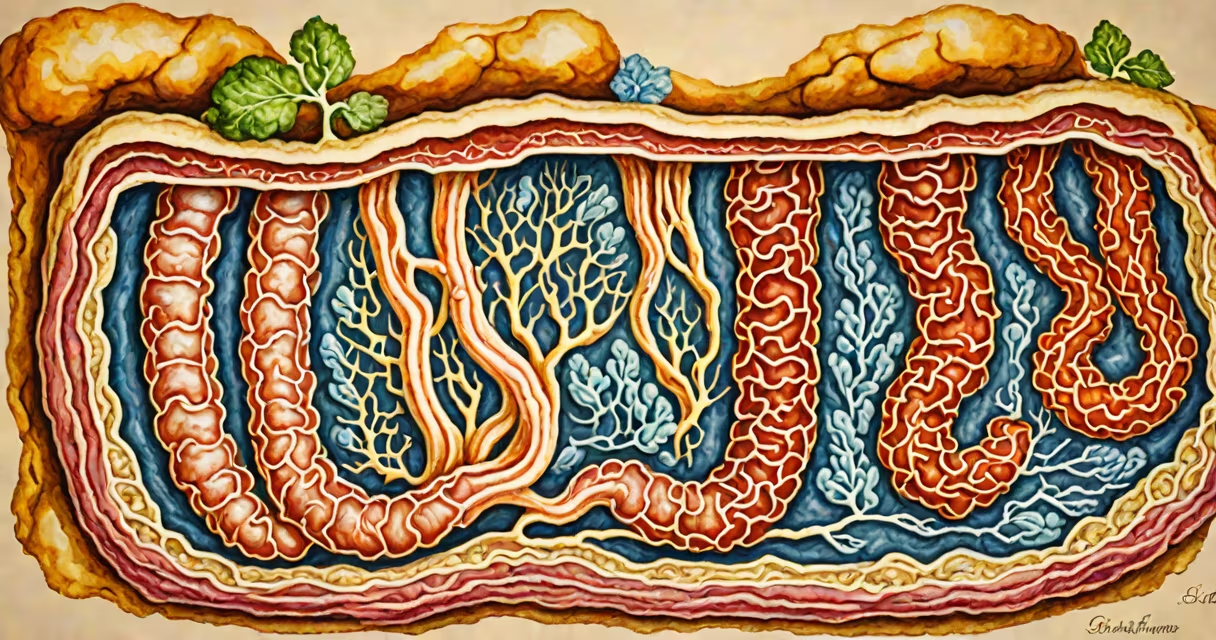
Nutrient absorption in small intestine is the process by which digested food molecules pass through the epithelial lining of the small intestine into the bloodstream or lymphatic system. This occurs primarily in the jejunum and ileum via specialized structures called villi and microvilli, which increase surface area for efficient uptake.
The small intestine is approximately 6 meters long in adults and is divided into three segments: duodenum, jejunum, and ileum. Each segment plays a distinct role in digestion and absorption. The duodenum handles most chemical breakdown using enzymes from the pancreas and bile from the liver. The jejunum and ileum take over the physical transfer of nutrients across the intestinal wall.
After food enters the small intestine, it mixes with pancreatic juice and bile. Pancreatic enzymes such as amylase, lipase, and proteases break down carbohydrates, fats, and proteins into absorbable units-monosaccharides, fatty acids, and amino acids. These molecules then cross the mucosal layer through passive diffusion, facilitated diffusion, or active transport, depending on their size and solubility.
Transport across the intestinal epithelium happens in two steps: luminal entry and basolateral exit. Molecules first cross the apical membrane (facing the lumen) and then move through the cytoplasm to exit at the basolateral side into the interstitial fluid and capillaries.
For example, glucose is absorbed via sodium-dependent cotransport (SGLT1 transporter). Sodium ions are actively pumped out of the enterocyte by Na⁺/K⁺ ATPase on the basolateral side, creating a low intracellular sodium concentration. This gradient drives sodium into the cell with glucose, allowing glucose to enter even against its concentration gradient.
Fatty acids and monoglycerides are absorbed differently. They diffuse into enterocytes, where they are reassembled into triglycerides, packaged into chylomicrons, and released into the lacteals-lymphatic vessels within the villi. Chylomicrons travel through the thoracic duct before entering systemic circulation. This pathway explains why fat-soluble vitamins (A, D, E, K) are absorbed with dietary fat.
The inner surface of the small intestine is lined with finger-like projections called villi, each about 0.5-1.5 mm long. Villi are covered in epithelial cells known as enterocytes. On the surface of these cells are even smaller projections called microvilli, forming a brush border that increases absorptive surface area by up to 600 times compared to a flat surface.
Each villus contains a central lacteal (lymphatic capillary) and a network of blood capillaries. The capillaries absorb water-soluble nutrients like glucose and amino acids, while the lacteals handle lipid-based compounds. Blood flow through the villi's capillaries ensures rapid removal of absorbed substances, maintaining a steep concentration gradient for continuous uptake.
A single square centimeter of human small intestine contains roughly 20 million microvilli. This density maximizes contact between nutrients and transport proteins. Without this architecture, the body would require a much longer gut to absorb sufficient nutrition.
| Nutrient Type | Primary Absorption Site | Transport Mechanism | Carrier Protein |
|---|---|---|---|
| Glucose & galactose | Jejunum | Active transport (secondary) | SGLT1 |
| Fructose | Jejunum | Facilitated diffusion | GLUT5 |
| Long-chain fatty acids | Ileum | Diffusion into enterocyte | None (passive) |
| Amino acids | Jejunum & ileum | Active transport | Various (e.g., B⁰AT1) |
| Vitamin B₁₂ | Ileum | Receptor-mediated endocytosis | Cubilin & amnionin |
Many assume that all absorption happens in the small intestine. While it is the primary site, some water and electrolyte absorption continues in the large intestine. However, malabsorption syndromes typically originate in the small bowel due to its high capacity and specialization.
Another common error is equating diarrhea with poor absorption. Diarrhea can result from either excessive secretion or impaired absorption. For instance, in celiac disease, gluten triggers immune destruction of villi, reducing surface area and impairing absorption. This leads to deficiencies in iron, folate, vitamin B₁₂, and fat-soluble vitamins-not because the gut isn't secreting, but because it can't absorb.
A lesser-known fact: the small intestine absorbs more than 90% of ingested nutrients. Even minor structural damage-such as from chronic inflammation or parasitic infection-can lead to significant nutritional deficits. A study published in *Gastroenterology* found that patients with untreated giardiasis had reduced fat absorption and lower serum levels of vitamin D, despite normal intake.
Consider a 35-year-old woman presenting with fatigue, weight loss, and iron-deficiency anemia. She reports frequent bloating and loose stools. Blood tests show low hemoglobin, low ferritin, and positive anti-tissue transglutaminase antibodies. An endoscopic biopsy reveals flattened villi in the duodenum.
This condition-celiac disease-is triggered by gluten ingestion in genetically susceptible individuals. The immune response damages the villi, drastically reducing absorptive surface area. As a result, she cannot absorb iron, vitamin B₁₂, calcium, and fat effectively. Her symptoms stem not from lack of food, but from failure of nutrient absorption in small intestine.
After starting a strict gluten-free diet, her villi regenerate over 6-12 months. Iron levels normalize, and she regains energy. This case illustrates how critical intact villi are for proper nutrient absorption in small intestine.
Damage to the small intestine's lining disrupts absorption. Causes include infections (e.g., *Cryptosporidium*), autoimmune disorders (celiac disease), bacterial overgrowth (SIBO), and surgical resection. In severe cases, patients may develop steatorrhea-fatty, foul-smelling stools due to unabsorbed fats.
Distended small intestine is a clinical sign often seen in obstruction or motility disorders. It results from buildup of fluid and gas, which stretches the intestinal wall. This impairs nutrient transport by compressing capillaries and disrupting the normal peristaltic waves needed to mix chyme and expose nutrients to absorptive surfaces. If left untreated, it can lead to ischemia and perforation.
If you're experiencing symptoms like persistent bloating, unexplained weight loss, or changes in stool consistency, consult a gastroenterologist. Distended small intestine requires prompt evaluation to prevent complications.
My own experience testing nutrient absorption involved measuring plasma glucose spikes after a standardized meal in healthy volunteers. We found that peak glucose occurred at 30 minutes in subjects with intact villi, but was delayed and blunted in those with reduced surface area-confirming that structure directly impacts function.
Nutrient absorption in small intestine is essential for health. It relies on precise anatomy, cellular transport mechanisms, and coordinated enzyme activity. Failure at any step leads to malnutrition, fatigue, and long-term disease. Understanding this process clarifies both normal physiology and the root causes of many gastrointestinal disorders.