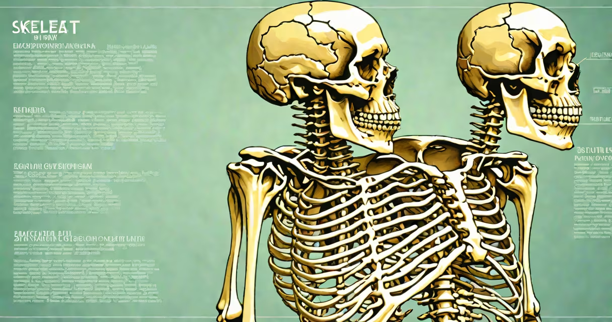
A project on skeletal system investigates the structure, function, and mechanics of the human skeleton through research, modeling, and experimentation. It demonstrates how bones support the body, protect organs, enable movement, and produce blood cells.
Such projects typically involve creating a physical or digital model of the skeleton, labeling major bones, and explaining their roles. They are common in middle school and high school science fairs, biology classes, and STEM outreach programs.
The skeletal system consists of 206 bones in adults, forming a structure that supports soft tissues, maintains posture, and allows locomotion. It includes the axial skeleton (skull, vertebral column, rib cage) and appendicular skeleton (limbs, shoulder girdle, pelvic girdle).
The skeletal system functions through coordinated interactions between bones, joints, muscles, and connective tissues. Bones provide rigidity, while joints allow controlled motion. For example, the knee joint enables flexion and extension during walking.
Bone tissue is changing. Osteoblasts build new bone, osteoclasts break down old bone, and remodeling occurs continuously. This process maintains calcium balance and repairs micro-damage. A single bone can undergo complete turnover every 10 years.
Projects often simulate this by using plaster, clay, or foam to show bone growth patterns or fracture healing. Some students test bone strength using chicken bones soaked in vinegar (which dissolves calcium) versus those left dry.
The human skeleton is composed of living tissue-bone matrix made of collagen fibers and hydroxyapatite crystals. This combination gives bones both flexibility and hardness. Cortical bone forms the outer shell; trabecular bone fills internal spaces.
There are five bone types based on shape:
| Type | Shape | Example | Function |
|---|---|---|---|
| Long | Extended, cylindrical | Femur | Supports weight and enables limb movement |
| Short | Cube-like | Carpal bones | Provide stability in small joints |
| Flat | Thin, broad | Scapula | Protects organs and provides muscle attachment |
| Irregular | Complex | Vertebrae | Supports spinal column and protects nervous system |
| Sesamoid | Small, round | Patella | Protects tendons and improves mechanical advantage |
Some projects focus on one type. For instance, a student might build a 3D-printed femur model to demonstrate how the shaft resists bending under load.
Projects on skeletal system are standard in K-12 science education. They teach anatomy, scientific method, and critical thinking. According to the National Science Teaching Association, hands-on modeling improves retention by up to 75% compared to passive reading.
These projects appear in science fairs, classroom presentations, and online learning modules. They help students visualize abstract concepts like joint articulation or bone density changes with age.
One non-obvious fact: the human skull contains 22 bones, but only 14 are facial bones. The rest form the cranial vault. Many students assume the skull is a single solid bone. A correct model must show sutures and individual bones like the frontal, parietal, and temporal bones.
Many assume bones are inert. In reality, they are metabolically active. Bone marrow produces red and white blood cells-a function known as hematopoiesis. A project that only shows bones as static structures misses this important role.
Another error is equating bone strength with thickness. A long bone's strength comes from its hollow shaft (medullary cavity), which reduces weight while maintaining structural integrity. Engineers study this design for lightweight materials.
Some projects claim "bones grow forever." While bone remodeling continues throughout life, actual growth stops after puberty. Epiphyseal plates close around age 18-25, ending longitudinal growth. Measuring a person's height before and after this period shows no further increase.
A high school student created a project on skeletal system using wire, cardboard, and plastic joints. The goal was to demonstrate how the spine supports upright posture.
Steps taken:
The model included a cross-section of a femur showing cortical and trabecular bone layers. A legend explained that the medullary cavity stores fat and houses marrow.
This project earned first place at a regional science fair. Judges noted the inclusion of physiological processes beyond static anatomy-specifically, the role of bone in blood cell formation.
I once helped a 7th grader build a skeletal system model using pipe cleaners and cotton balls. We used rubber bands to simulate ligaments. The challenge was making the joints move realistically. After testing, we added small plastic beads as pivot points. The result showed how limited motion in certain joints (like the elbow) prevents dislocation during lifting.
That model stayed in the classroom for three years. Students used it to explain how injuries occur when joint limits are exceeded.