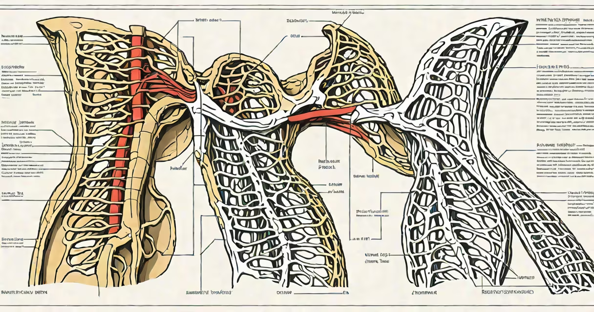
The thoracic duct is the primary conduit for lymph return from most of the body into the bloodstream, draining approximately 3 liters of lymph daily from the lower body, left upper body, and left side of the head and neck.
It originates in the cisterna chyli, a dilated sac located at the level of the first lumbar vertebra, formed by the union of the intestinal and lumbar lymphatic trunks. From there, it ascends through the posterior mediastinum, passing behind the esophagus and anterior to the vertebral column. It pierces the diaphragm at the aortic hiatus and continues upward through the neck, terminating at the junction of the left internal jugular and left subclavian veins.
At its termination, the thoracic duct empties lymph into the venous system via a valve-controlled entry, preventing backflow. This process maintains fluid balance, supports immune surveillance, and transports dietary lipids absorbed from the small intestine via chylomicrons.
Lymph enters the thoracic duct through numerous collecting vessels that drain interstitial fluid from tissues throughout the body. These vessels converge into larger trunks-intestinal, lumbar, bronchomediastinal, and subclavian-that feed into the cisterna chyli and then into the duct proper.
As lymph travels upward, it passes through a series of valves that prevent retrograde flow. These valves ensure unidirectional movement despite changes in intrathoracic pressure during breathing. The duct's wall contains smooth muscle fibers that contract rhythmically, aiding propulsion of lymph toward the venous system.
When the thoracic duct empties into the left subclavian vein, lymph mixes with blood. This returns proteins, immune cells, and excess fluid to circulation. Without this drainage, edema would develop rapidly in dependent tissues due to fluid accumulation.
The thoracic duct lies in close proximity to important organs and structures. In the abdomen, it runs alongside the aorta and inferior vena cava. In the thorax, it courses between the azygos vein and the esophagus, often lying within the posterior mediastinum.
In the neck, the duct ascends along the left side of the trachea and lies beneath the sternocleidomastoid muscle. Its terminal portion is typically near the left brachiocephalic vein, making it vulnerable during surgical procedures involving the neck or upper chest.
Understanding spatial relationships is critical in surgery. For example, during a left-sided thoracotomy, surgeons must identify the duct to avoid injury. Damage can result in chylothorax-a condition where chyle leaks into the pleural cavity, causing respiratory distress and malnutrition.
| Anatomical Segment | Location | Drainage Area | Clinical Significance |
|---|---|---|---|
| Cisterna chyli | L1 vertebra, posterior abdominal wall | Intestinal and lumbar lymph | Site of lymph collection; site of potential trauma in abdominal surgery |
| Abdominal segment | Through posterior mediastinum | Lower limbs, abdomen, left upper body | Common site of lymph node metastasis in cancer staging |
| Thoracic segment | Posterior mediastinum, between esophagus and azygos vein | Left lung, heart, left arm | High risk during cardiac or thoracic surgery |
| Neck segment | Left side of neck, near carotid sheath | Left head and neck, left upper limb | Site of iatrogenic injury during thyroidectomy |
Many assume the thoracic duct drains both sides of the body equally. This is incorrect. The right lymphatic duct handles the right upper quadrant (right arm, right side of head and neck), while the thoracic duct manages everything else.
Another misconception is that the thoracic duct only carries clear lymph. In reality, it transports chyle-milky white fluid rich in triglycerides-after fat absorption in the small intestine. This explains why chylothorax appears cloudy and lipid-rich under microscopy.
A lesser-known fact: the thoracic duct may have variable anatomy. In about 10% of people, it enters the venous system at the junction of the right subclavian and internal jugular veins. This variation is clinically relevant during imaging interpretation and surgical planning.
A 62-year-old man underwent a left thyroid lobectomy for a benign nodule. Postoperatively, he developed dyspnea and decreased breath sounds on the left. A chest X-ray showed a left-sided pleural effusion. Thoracentesis revealed fluid with high triglyceride levels-diagnostic of chylothorax.
Imaging confirmed a leak from the thoracic duct near the left jugular vein. The patient was managed conservatively with a low-fat diet and medium-chain triglyceride supplementation. After 14 days, the effusion resolved. Surgical ligation of the duct was avoided.
This case illustrates how precise knowledge of thoracic duct anatomy prevents complications. Surgeons must recognize the duct's location relative to the recurrent laryngeal nerve and inferior thyroid artery to avoid injury.
Errors in identifying the thoracic duct during surgery lead to significant morbidity. Chylothorax occurs in 0.5% to 1% of thyroidectomies and requires prolonged hospitalization and nutritional support.
Medical students and clinicians must distinguish the thoracic duct from the right lymphatic duct. The right duct is shorter, ends in the right subclavian vein, and drains only the right upper body. Confusing the two leads to misinterpretation of lymphatic drainage patterns in diagnostic imaging.
Thoracic duct anatomy is not merely a memorization task-it is a functional blueprint for fluid homeostasis, immunity, and surgical safety. Its structure reflects evolutionary design for efficient return of filtered fluid to the circulatory system.