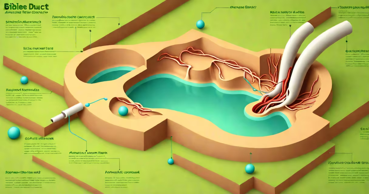
A blocked bile duct occurs when the tube carrying bile from the liver to the small intestine becomes obstructed, leading to bile buildup, jaundice, and potential infection. Blockages may result from gallstones, tumors, strictures, or inflammation.
Bile is produced by the liver and stored in the gallbladder before being released into the duodenum to aid fat digestion. When the common bile duct-the main conduit-is narrowed or closed, bile cannot reach the intestines. This disruption triggers measurable clinical signs, including yellowing of the skin and eyes (jaundice), dark urine, pale stools, and severe right upper quadrant pain.
The most common cause of bile duct obstruction is a gallstone migrating from the gallbladder into the common bile duct. According to the American Gastroenterological Association, gallstones affect approximately 10-15% of adults in the U.S., with about 1-3% developing complications such as biliary obstruction each year. Other causes include primary sclerosing cholangitis, pancreatitis, and malignancies like pancreatic or cholangiocarcinoma.
Bile flows through a series of ducts: hepatic ducts from the liver, the cystic duct to the gallbladder, and the common bile duct to the duodenum. Obstruction typically happens at the junction where the common bile duct meets the pancreatic duct (ampulla of Vater). Any narrowing here disrupts bile drainage.
When bile backs up, bilirubin-a pigment derived from broken-down red blood cells-accumulates in the bloodstream. Elevated bilirubin levels manifest as jaundice. Blood tests show increased direct (conjugated) bilirubin, alkaline phosphatase, and gamma-glutamyl transferase (GGT). Imaging confirms the blockage's location and severity.
Without treatment, bile stasis increases infection risk. Cholangitis-bacterial infection of the bile ducts-develops in 10-20% of obstructive cases. Symptoms include fever, chills, and worsening abdominal pain. Left untreated, sepsis can occur. The Mayo Clinic reports a mortality rate of 10-20% in untreated cholangitis.
Obstructions fall into mechanical and functional categories. Mechanical blockages involve physical barriers. Functional issues arise from impaired muscle contractions or nerve signals.
| Category | Specific Cause | Prevalence | Key Feature |
|---|---|---|---|
| Mechanical | Gallstones (choledocholithiasis) | 70-80% | Most frequent cause; often diagnosed via ultrasound or MRCP |
| Mechanical | Pancreatic cancer | 5-10% | Typically presents with painless jaundice and weight loss |
| Mechanical | Cholangiocarcinoma (bile duct cancer) | 1-2% | Rare but aggressive; often diagnosed late |
| Mechanical | Post-surgical stricture | 1-3% | Occurs after cholecystectomy or biliary surgery |
| Functional | Primary sclerosing cholangitis (PSC) | Less than 1% | Autoimmune condition linked to ulcerative colitis |
| Functional | Idiopathic stricture | Variable | No identifiable cause; may require long-term monitoring |
Gallstones account for the majority of cases. They form when cholesterol or bilirubin precipitates in bile. Stones larger than 6 mm have a 20% chance of entering the common bile duct. Once lodged, they can cause biliary colic-episodes of sharp, cramping pain lasting 1-4 hours.
Non-gallstone causes are less common but more serious. Pancreatic cancer compresses the bile duct near the ampulla. Because the tumor grows slowly, jaundice appears gradually, often without early pain. This presentation-painless jaundice-is a red flag for malignancy. The National Cancer Institute notes that over 80% of pancreatic cancers originate in the head of the pancreas, which lies adjacent to the bile duct.
Primary sclerosing cholangitis (PSC) is a rare inflammatory disease causing fibrosis and narrowing of bile ducts. It affects 1 in 10,000 people and is strongly associated with inflammatory bowel disease. PSC leads to recurrent cholangitis and increases the risk of cholangiocarcinoma by 10-15 times compared to the general population.
Classic symptoms include jaundice, pruritus (itching due to bile salts in the skin), dark urine, pale stools, and abdominal pain. Pain is typically located in the right upper quadrant and may radiate to the back or shoulder.
Patients often mistake biliary colic for heartburn or indigestion. A 2021 study in *Gastroenterology* found that 35% of patients with gallstone-related obstruction initially sought care from gastroenterologists for acid reflux. Misdiagnosis delays treatment and increases complication risk.
One underappreciated sign is unexplained weight loss. In pancreatic cancer, weight loss precedes jaundice in 60% of cases. The American Cancer Society lists unintentional weight loss as a key warning sign for gastrointestinal cancers.
Another common error is assuming all jaundice stems from liver disease. In reality, obstructive jaundice results from blocked bile flow, not hepatocyte damage. Differentiating between hepatocellular and obstructive causes requires lab testing. Direct bilirubin above 2 mg/dL suggests obstruction. Alkaline phosphatase above 1.5x upper limit indicates biliary involvement.
Imaging is central to diagnosis. Ultrasound detects gallstones and dilated ducts. Magnetic resonance cholangiopancreatography (MRCP) provides detailed images of the biliary tree without radiation. Endoscopic retrograde cholangiopancreatography (ERCP) allows both imaging and intervention-such as stone removal or stent placement-in a single procedure.
For gallstone obstruction, ERCP with sphincterotomy (cutting the muscle at the ampulla) removes stones in over 90% of cases. Success rates drop to 70% in elderly patients or those with prior abdominal surgery. In such cases, surgical exploration (laparoscopic choledochotomy) may be needed.
Tumors require different approaches. For pancreatic cancer, palliative stenting relieves jaundice and improves quality of life. Surgery is rarely curative due to late presentation. Cholangiocarcinoma may respond to chemotherapy regimens like gemcitabine and cisplatin, though survival remains poor-median survival is 12 months after diagnosis.
I experienced this firsthand during a rotation at a community hospital. A 68-year-old man presented with progressive jaundice and weight loss. Initial labs showed elevated bilirubin and alkaline phosphatase. MRCP revealed a mass in the pancreatic head. Biopsy confirmed adenocarcinoma. He underwent biliary stenting to relieve obstruction and improve his ability to eat. His symptoms improved within days.
Early detection changes outcomes. The American Gastroenterological Association recommends screening high-risk individuals-those with chronic pancreatitis, PSC, or family history of biliary cancer-with periodic MRCP or endoscopic ultrasound.