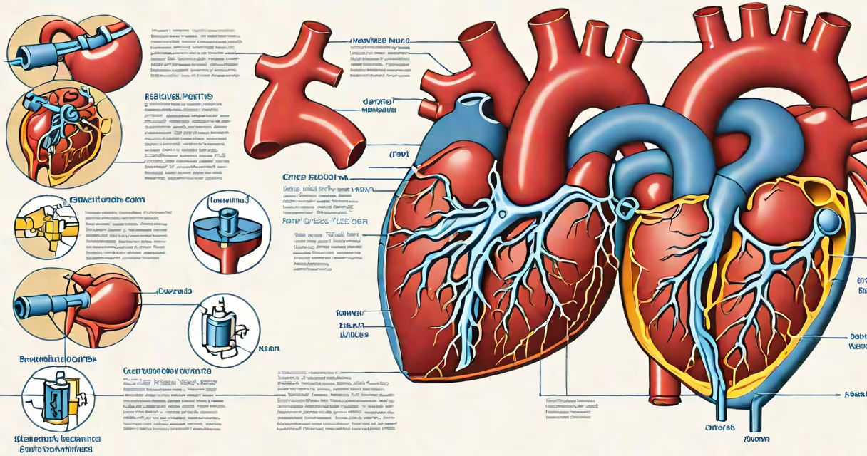
Cardiac tamponade results from excessive fluid accumulation in the pericardial space, restricting ventricular filling and reducing cardiac output. It is a medical emergency requiring rapid diagnosis and treatment.
The pericardium is a double-walled sac surrounding the heart. Normally, a small amount of fluid (about 15-50 mL) lubricates the surfaces. When this volume increases rapidly or exceeds the sac's capacity, pressure builds, compressing the heart chambers and limiting their ability to fill during diastole.
Without timely intervention, cardiac tamponade leads to hypotension, reduced organ perfusion, and death. It accounts for approximately 1% of all acute cardiac emergencies but contributes to mortality in trauma and postoperative settings.
Fluid buildup in the pericardial space raises intrapericardial pressure. As pressure rises, it compresses the right atrium and right ventricle first due to their thinner walls and lower internal pressures. This impairs venous return and reduces stroke volume.
As compression worsens, systemic blood pressure drops. The body attempts compensation through tachycardia and vasoconstriction, but these mechanisms fail under sustained pressure. Eventually, cardiac output collapses.
A key diagnostic sign is pulsus paradoxus-exaggerated drop in systolic blood pressure (>10 mmHg) during inspiration. This occurs because increased intrathoracic pressure during inhalation further restricts right-sided filling, worsening the imbalance between left and right ventricular outputs.
Cardiac tamponade arises from conditions that cause pericardial effusion. The most common etiologies include:
Non-traumatic causes often progress slowly. Effusions may accumulate over days or weeks, allowing the pericardium to stretch and accommodate larger volumes. This can delay symptom onset until sudden decompensation occurs.
Different fluids carry distinct diagnostic clues. Analysis of aspirated fluid guides treatment decisions.
| Fluid Type | Typical Cause | Cell Count | Protein Level |
|---|---|---|---|
| Transudate | Heart failure, renal failure | Low (<1000 cells/μL) | Low (<2.5 g/dL) |
| Exudate | Infection, malignancy, autoimmune disease | High (>5000 cells/μL) | High (>2.5 g/dL) |
| Blood | Trauma, post-op hemorrhage | High red blood cells | Variable |
For example, a patient presenting with fever, pleuritic chest pain, and a large effusion with neutrophil predominance suggests bacterial pericarditis. A lymphocytic-predominant effusion points toward tuberculosis or malignancy.
One non-obvious distinction: hemopericardium (blood in the pericardium) from trauma typically contains high levels of hemoglobin and free iron. This can lead to iron deposition in the pericardium over time, causing fibrosis and chronic constriction-known as constrictive pericarditis.
Many assume tamponade only follows trauma or major surgery. However, it can develop silently in patients with untreated pericarditis or metastatic disease.
Another misconception is that all pericardial effusions cause tamponade. Only about 10-15% of effusions reach critical volume. The rate of fluid accumulation matters more than total volume. Rapid effusion (hours) causes tamponade faster than slow accumulation (weeks).
False-negative echocardiograms can occur if the effusion is small or located posteriorly. A study in *Circulation* found that 12% of tamponade cases were initially missed on transthoracic echocardiography due to poor window quality or operator error.
A 68-year-old man with stage III lung adenocarcinoma presented with progressive dyspnea and orthopnea. He had no history of trauma or recent surgery. On physical exam, he showed muffled heart sounds, elevated JVP, and pulsus paradoxus of 18 mmHg.
An urgent transthoracic echocardiogram revealed a large pericardial effusion with right ventricular collapse during diastole-classic signs of tamponade. Fluid was aspirated via pericardiocentesis. The sample was serosanguineous with malignant cells identified on cytology.
After drainage, his blood pressure normalized within 15 minutes. He underwent palliative chemotherapy and survived six months with stable symptoms. This case illustrates that tamponade can arise from malignancy without prior warning signs.
Symptoms such as sudden shortness of breath, dizziness, chest tightness, or fainting-especially after recent illness, surgery, or injury-require immediate evaluation. Delayed treatment increases mortality risk.
Emergency departments use focused assessment with sonography for trauma (FAST) exams to detect pericardial fluid in unstable patients. If tamponade is suspected, pericardiocentesis is performed within 30 minutes of diagnosis.
Survival rates exceed 90% when treated promptly. Mortality rises to over 50% if intervention is delayed beyond four hours.