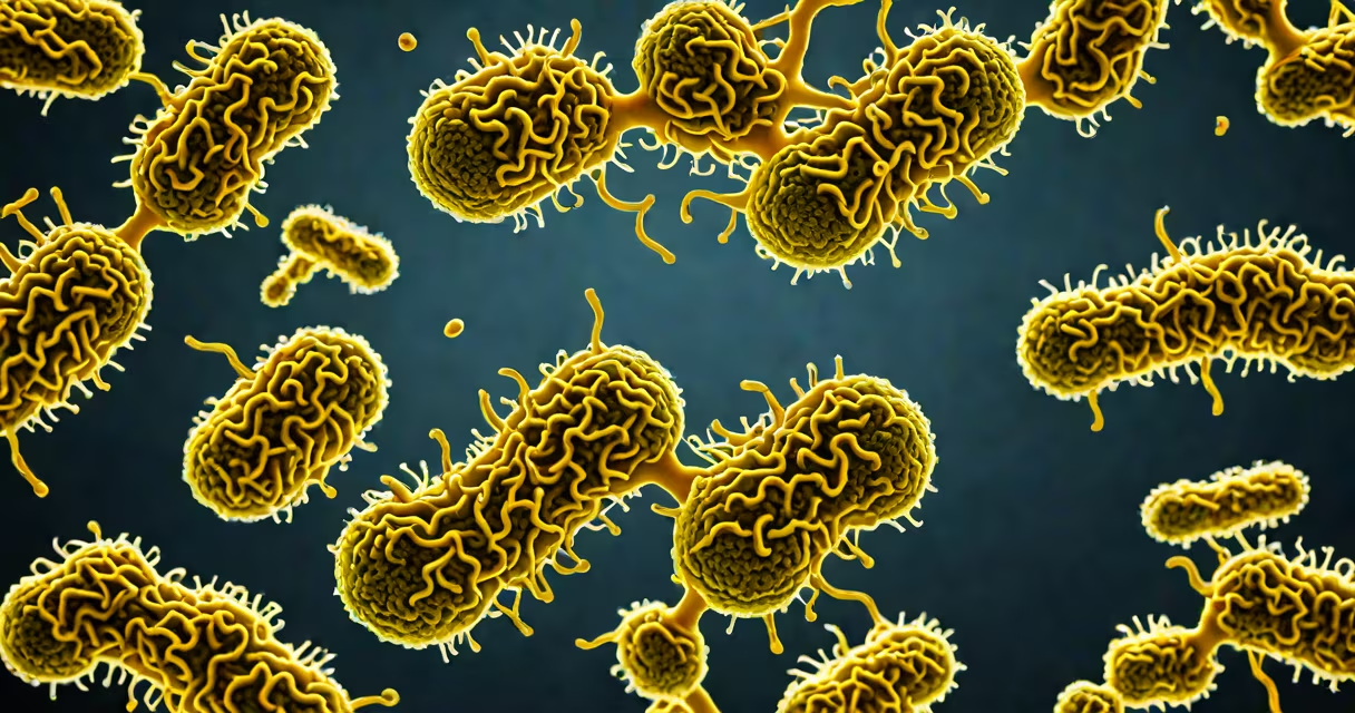
Shiga toxin 1 and 2 are highly toxic proteins secreted by specific pathogenic strains of Escherichia coli, most E. coli O157:H7, that damage intestinal cells and trigger life-threatening complications such as hemolytic uremic syndrome. These toxins inhibit protein synthesis in human cells by cleaving a single adenine residue in the 28S ribosomal RNA of eukaryotic ribosomes.
Shiga toxin 1 (Stx1) and Shiga toxin 2 (Stx2) are structurally related but distinct. Both are AB5 toxins-composed of one enzymatically active A subunit and five binding B subunits-but differ in amino acid sequence, potency, and biological effects. Stx1 is nearly identical to the toxin produced by Shigella dysenteriae, the bacterium that causes shigellosis. Stx2 is more commonly associated with severe disease outcomes, including renal failure and neurological symptoms.
The toxins are encoded by bacteriophages integrated into the E. coli genome. When the phage replicates, it triggers toxin production. This means toxin expression is not constant-it occurs during bacterial lysis or under stress conditions. Once released, the B subunits bind to globotriaosylceramide (Gb3) receptors on endothelial cells, particularly in the kidneys and brain. The A subunit then enters the cell and halts protein synthesis, leading to cell death.
Each Shiga toxin molecule consists of one A subunit and five B subunits arranged in a pentamer. The B subunits recognize and bind to Gb3, a glycolipid receptor abundant on the surface of human glomerular endothelial cells and neurons. After binding, the complex undergoes endocytosis and traffics retrograde through the Golgi apparatus to the endoplasmic reticulum.
In the ER, the A subunit is cleaved and translocated into the cytosol. There, it acts as an N-glycosidase, removing a single adenine base from the sarcin-ricin loop of the 28S rRNA in the 60S ribosomal subunit. This modification irreversibly inactivates the ribosome, preventing translation of mRNA into protein. Cells cannot produce essential proteins and die within hours.
A 2018 study published in *Nature Microbiology* demonstrated that Stx2 induces stronger inflammatory responses in human kidney cells than Stx1, partly due to enhanced activation of NF-κB signaling pathways. This heightened inflammation contributes to vascular damage and microthrombi formation, which are hallmarks of hemolytic uremic syndrome (HUS).
While Stx1 and Stx2 are the primary forms, several variants exist. Stx1 has two major subtypes: Stx1a and Stx1c. Stx2 has five known subtypes: Stx2a, Stx2b, Stx2c, Stx2d, and Stx2e. Stx2a is the most frequently isolated from patients with HUS and is strongly linked to severe disease.
| Toxin Type | Origin | Potency (relative) | Association with HUS | Key Structural Difference |
|---|---|---|---|---|
| Stx1a | E. coli O157:H7, Shigella dysenteriae | 1.0 | Moderate | Identical to Shigella toxin |
| Stx2a | E. coli O157:H7, O104:H4 | 1.5-2.0 | High (most common in HUS cases) | Higher affinity for Gb3, increased stability |
| Stx2c | E. coli O111, O103 | 1.2 | High | More resistant to heat and pH changes |
| Stx2d | E. coli O157:H7 (rare) | 0.8 | Mild | Lower binding affinity |
Stx2a is responsible for the majority of HUS cases in North America and Europe. A 2020 CDC report found that 78% of HUS cases in a 10-year surveillance period were linked to Stx2a-producing E. coli. Stx1 is less likely to cause HUS but still contributes to severe diarrhea and bloody stools.
Shiga toxins are not used therapeutically. Their presence in nature serves a role in bacterial survival and host invasion. By damaging intestinal epithelium, they disrupt the mucosal barrier, allowing E. coli to access deeper tissues and nutrients. Toxin-induced cell death also releases iron and other nutrients from lysed cells, supporting bacterial growth.
Once in the bloodstream, Stx2 targets capillaries in the kidneys and brain. Glomerular endothelial cells express high levels of Gb3, making them vulnerable. Damage leads to thrombotic microangiopathy-small blood clots form in vessels, destroying red blood cells and impairing kidney function. This cascade results in acute kidney injury, hemolytic anemia, and low platelet counts-diagnostic features of HUS.
Neurological complications occur when Stx crosses the blood-brain barrier or damages cerebral endothelium. Seizures, altered mental status, and coma have been documented in pediatric HUS cases. A 2019 study in *The Journal of Infectious Diseases* showed that Stx2a was detected in cerebrospinal fluid samples from two children with neurologic HUS, confirming direct CNS involvement.
Many assume all E. coli strains produce Shiga toxin. This is false. Only a subset of E. coli-specifically enterohemorrhagic E. coli (EHEC)-produce Stx1 and Stx2. Non-pathogenic E. coli strains, such as those in the gut microbiome, do not carry the genes.
Another misconception is that antibiotics treat Shiga toxin infection. They do not. In fact, antibiotic use increases the risk of HUS. A 2016 meta-analysis in *Clinical Infectious Diseases* found that patients treated with fluoroquinolones had a 2.3-fold higher incidence of HUS compared to untreated controls. Antibiotics promote phage induction, increasing toxin release and systemic exposure.
Some believe toxin detection requires culturing bacteria. Modern diagnostics rely on PCR testing for stx1 and stx2 genes in stool samples. A 2021 study in *Emerging Infectious Diseases* reported that PCR-based detection identified Stx2 in 94% of confirmed HUS cases, while culture missed 30% due to low bacterial load.
In May 2011, Germany experienced a large outbreak of E. coli O104:H4 infections. Over 4,000 people were infected, and 53 died. The strain produced Stx2c and Stx2a, both of which contributed to widespread HUS. Unlike typical E. coli O157:H7, this strain had acquired a pathogenicity island containing the stx2 gene and additional virulence factors.
Patients presented with severe bloody diarrhea, followed by rapid onset of kidney failure. In intensive care units, 90% of HUS patients required dialysis. The outbreak highlighted that Stx2 variants beyond Stx2a can cause severe disease and that non-O157 strains are increasingly significant in public health.
I observed a diagnostic lab process during a training rotation at a state public health department. A stool sample from a child with bloody diarrhea was tested using a multiplex PCR assay targeting stx1, stx2, and eae genes. The result showed positive signals for both stx1 and stx2. The patient was hospitalized, developed HUS within 48 hours, and required plasma exchange therapy. The lab's ability to detect Stx2 rapidly guided treatment decisions.
When you suspect Shiga toxin infection, especially in children with bloody diarrhea, test for stx1 and stx2 genes. Avoid antibiotics. Focus on hydration and supportive care. If HUS develops, consult nephrology early.
Understanding shiga toxin 1 and 2 is critical for diagnosing and managing E. coli-related illnesses. These toxins are not interchangeable-Stx2, particularly Stx2a, poses a greater threat than Stx1. Their mechanism of action involves targeted ribosomal destruction, leading to cellular collapse and organ failure. Early detection and avoidance of antibiotics improve outcomes.