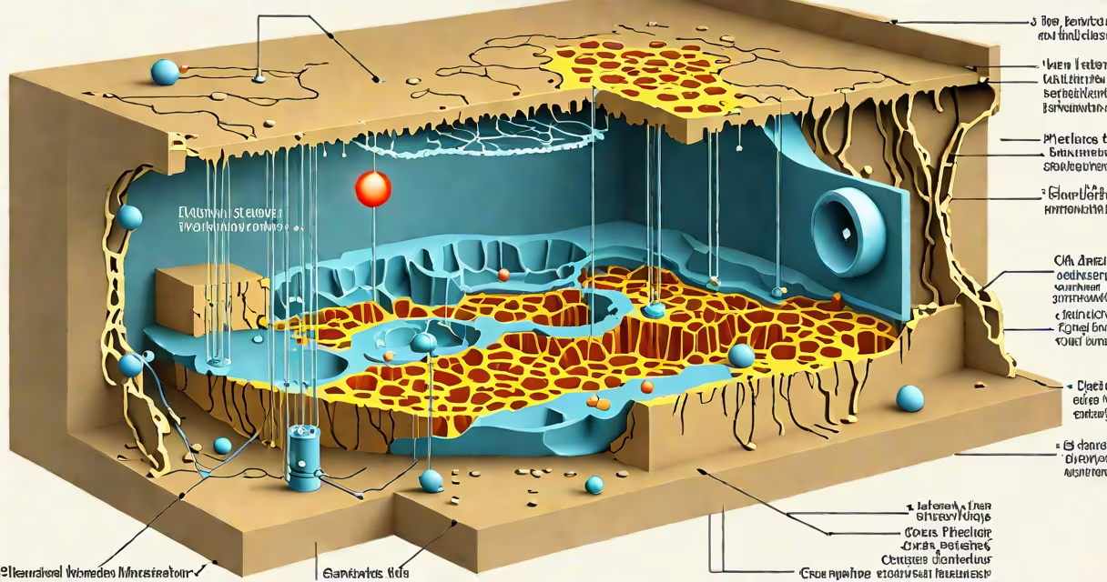
The plasma membrane is located at the outer boundary of every animal and plant cell, forming a continuous lipid bilayer that separates the cell's internal environment from the extracellular space.
This membrane defines the cell's perimeter and regulates what enters and exits. It is not a static wall but a changing interface shaped by proteins, cholesterol, and glycolipids embedded within its structure.
Its position is consistent across eukaryotic cells: immediately adjacent to the cytoplasm on the inner side and in direct contact with the extracellular matrix or surrounding fluid on the outer side. In animal cells, it lies just beneath the glycocalyx-a sugar-coated layer that aids in cell recognition and protection.
The plasma membrane maintains homeostasis by controlling ion flow, nutrient uptake, and waste removal. It uses selective permeability-allowing small, nonpolar molecules like oxygen and carbon dioxide to pass freely while blocking larger or charged substances unless aided by transport proteins.
For example, glucose cannot cross the membrane unassisted. Instead, it binds to GLUT4 transporter proteins embedded in the membrane, which change shape to shuttle the molecule into the cell. This mechanism is essential in muscle and fat cells during insulin response.
Without the plasma membrane's precise positioning, cellular integrity collapses. A damaged membrane leads to uncontrolled leakage of potassium ions and influx of calcium, triggering apoptosis-the programmed death of the cell.
The plasma membrane is primarily composed of phospholipids arranged in a bilayer, with hydrophilic heads facing outward toward water-based environments and hydrophobic tails pointing inward. Cholesterol modulates fluidity, preventing the membrane from becoming too rigid or too fluid depending on temperature.
Proteins are integral to function. Transmembrane proteins span the entire width of the membrane and serve as channels, receptors, or enzymes. For instance, the epidermal growth factor receptor (EGFR) sits in the plasma membrane and activates signaling pathways when bound to its ligand.
Carbohydrates attach to lipids (glycolipids) or proteins (glycoproteins) on the extracellular surface, forming the glycocalyx. These sugars act as identity markers-blood type AB is determined by specific glycoproteins on red blood cell membranes.
Many confuse the plasma membrane with the cell wall. The cell wall exists only in plants, fungi, and bacteria-it provides structural support but is not selectively permeable. In contrast, the plasma membrane controls molecular traffic.
Another error is assuming the membrane is uniform. It is highly asymmetric: the inner leaflet contains phosphatidylethanolamine and phosphatidylserine, while the outer leaflet is rich in phosphatidylcholine and sphingomyelin. This asymmetry is maintained by flippase, floppase, and scramblase enzymes.
A lesser-known fact: the plasma membrane is not a flat surface. It forms microdomains called lipid rafts-regions enriched in cholesterol and sphingolipids that cluster signaling proteins. These rafts are involved in immune responses and viral entry, such as HIV using them to infect T cells.
Different cell types have specialized membrane compositions based on function:
| Cell Type | Membrane Specialization | Functional Purpose |
|---|---|---|
| Nerve cell (neuron) | High density of voltage-gated sodium channels | Generates action potentials for signal transmission |
| Red blood cell | Abundant band 3 protein and spectrin network | Maintains biconcave shape and flexibility for capillary passage |
| Intestinal epithelial cell | Microvilli with brush border enzymes | Increases surface area for nutrient absorption |
These adaptations show that while the plasma membrane's basic structure is conserved, its functional complexity varies widely.
In 2018, researchers at the University of California, San Diego, studied how muscle cells repair their plasma membrane after mechanical stress. They found that dysferlin-a protein normally stored in intracellular vesicles-rapidly migrates to the site of membrane rupture within seconds.
Dysferlin acts like a patch: it fuses vesicles to the damaged area, sealing the breach. Mutations in the dysferlin gene cause limb-girdle muscular dystrophy type 2B, a condition where muscle cells repeatedly lose membrane integrity due to failed repair.
This demonstrates that the plasma membrane's location is not passive-it actively participates in cellular defense through changing remodeling.
While the plasma membrane is the outermost boundary, other organelles have their own membranes. The nuclear envelope, mitochondria, lysosomes, and endoplasmic reticulum each have distinct lipid compositions and protein sets tailored to their roles.
For example, mitochondrial membranes are rich in cardiolipin-a phospholipid not found in the plasma membrane. Cardiolipin stabilizes electron transport chain complexes and is essential for ATP production.
Confusing these membranes leads to errors in interpreting disease mechanisms. In Barth syndrome, mutations in the tafazzin enzyme disrupt cardiolipin remodeling, causing heart failure and muscle weakness. This is not a defect in the plasma membrane but in an internal organelle membrane.
I once observed live zebrafish embryos under a confocal microscope. Fluorescently labeled antibodies tagged the plasma membrane of developing neurons. As axons extended, the membrane visibly bulged and reshaped itself-forming filopodia and lamellipodia-driven by actin polymerization beneath the membrane.
Seeing the membrane dynamically respond to guidance cues confirmed that it is not a barrier but a responsive interface. Its location is not fixed-it adapts in real time to cellular needs.
The plasma membrane is not just a boundary-it is a functional interface. Its location at the cell's edge enables communication, transport, and protection. It is the site of hormone reception, nutrient uptake, and immune recognition.
To understand where the plasma membrane is, consider that it is both everywhere and nowhere-it defines the cell's limits while being invisible to the naked eye. Its position is fundamental to life as we know it.